fbpx
bicmd

Expert neck, back, & spine care at your fingertips

You’re tired of living with chronic pain, but you want to avoid any unnecessary procedures. With access to the country’s best experts, you can get back to doing the activities you love.

years

average experience of BICMD neck, back, & spine expert

%

of neck, back, & spine conditions covered

minutes

average 1:1 time spent with a BICMD specialist during encounter

Feel completely confident in your journey toward neck, back, & spine health

Whether you need a second opinion or you’re just starting out, it’s time to work with a specialist at Best In Class MD. Find your condition or recommended treatment below and see how we can help.

Back Pain Overview

Back pain is one of the most common reasons individuals visit a doctor. In fact, it is estimated that 90% of people world-wide experience back pain at some point in their life. Half of the world’s population experience back pain at least once a year. Chronic back pain is not always caused by an underlying disease, but can be from overuse, too much exercise, sleeping in an uncomfortable positions and other muscle-related reasons. Back pain can come and go, but when it becomes severe and interrupts daily activities, it is time to seek an orthopedic spine specialist. At BICMD our “Best in Class” surgeons and specialists can give you an expert diagnosis as well as treatment options through our orthopedic telemedicine platform. If you are looking for a second opinion, you have found the best doctors in the industry, here at BICMD and can use our state-of-the art telemedicine platform for your back pain options.

What is the difference between chronic and acute back pain?

Back pain is classified by either chronic or acute. Acute back pain is a strain or sprain of the soft tissues in the back. These can include muscles, tendons and ligaments. Acute back pain can occur after lifting a heavy object incorrectly or over-exercising; it is considered acute when the pain lasts for less than a month. Chronic back pain is pain that has lasted more than three months and can be caused by any number of injuries or conditions. Individuals who have back pain that lasts more than three days, especially if severe, needs to see a physician or consult with one of our experts at BICMD.

What causes chronic back pain?

Any number of problems can affect the back due to its complex structure. Chronic neck and back pain can be caused by:

  • Tendonitis or tendinopathy – Both cause back and neck pain: Tendonitis is the inflammation of the tendon. Tendinopathy is a degeneration of the collagen protein that forms the actual tendon.
  • Osteoarthritis
  • Degenerative Conditions including disc disease  – The gradual loss of normal structure and function of the spine over time.
  • Herniated or bulging disc
    • Cervical disc herniation – Can cause neck pain and pain in the shoulders, shoulder blade and even numbness and tingling in the hands arms.
    • Lumbar disc herniation  – Causes pain in the lower back, buttocks and can radiate pain down one or both legs. Severe lumbar disc herniation that causes foot-drop will require immediate medical care.
  • Spinal Stenosis
    • Lumbar Spinal Stenosis – Narrowing of the spinal canal which can put pressure on the nerves and spinal cord.
    • Cervical Spinal Stenosis – Myelopathy Narrowing of the spinal canal in the neck, if left untreated, can result in Myelopathy or an injury to the spinal cord.
  • Scoliosis – A “C” or “S” curve in the spine.
  • Spinal cord injury

How is acute and chronic back pain diagnosed?

The back pain experts at BICMD begin the diagnosis for acute and chronic back pain with a thorough patient history. The patient’s symptoms and their severity will help determine the diagnostic testing that may be needed.  One or more of the following tests can help properly diagnose your back pain:

  • MRI – Magnetic Resonance Imaging – Uses strong magnetic fields and radio waves to see detailed images inside the body.
  • CT Scan – Computerized Tomography – Uses computers and a rotating x-ray machine to see cross-sections within the body.
  • X-ray – Shows the bones and can be a “first step” in diagnosing the cause of back pain

What is the treatment for acute and chronic back pain?

The recommended treatment for acute and chronic back pain will vary greatly depending upon symptoms, severity and if there is a spinal condition that needs to be addressed. In general, acute back pain does not need surgery and most cases resolve relatively quickly. Patients are advised to rest, but only for a day or two. It is not recommended that patients remain in bed, as it can make symptoms worse. Over the counter pain medication, muscle relaxers, hot and cold therapy, or working with a physical therapist can help relieve pain.

Back pain that is chronic or has an underlying condition, such as a herniated disc or degenerative joint disease have varying treatment options. It is important to obtain an expert opinion from one of our “Best in Class” back and spine experts at BICMD before undergoing any kind of surgical treatment. Depending on your diagnosis, they may recommend one of the following treatments for chronic back pain:

  • Spinal Injections  – Highly effective for immediate relief of back pain.
    • Fluoroscopic/x-ray-guided epidural injections
    • Facet joint injections
    • Medial branch block – radiofrequency denervation
  • Sacroiliac joint injections
  • Disc Replacement
    • Cervical Disc Replacement – Replaces the damaged disc. New methods and materials maintain the flexibility of the cervical vertebrae.
  • Lumbar Laminectomy – Creates space in the lumbar canal of the spine and relieves pressure on the spinal cord.
  • Cervical Laminoplasty – Relieves pressure on the spinal cord in the neck by reshaping or repositioning the bones in the neck.
  • Lumbar Microdiscectomy
  • Lumbar Fusion – Fuses two or more bones together and is designed to stop the motion of the painful vertebral segment.


For more information about chronic back pain and acute back pain as well as the different treatments you should consider, or to receive an expert consultation about your back and neck pain, please contact our specialists, by clicking on “Get Started”.  You will be connected to one of our orthopedic telemedicine experts through our state-of-the-art telemedicine platform.

What are degenerative conditions of the spine?

Degenerative conditions of the spine are sometimes called degenerative spine disease. It usually occurs as individuals age and involves the gradual loss of the normal function and structure of the spine over time. Degenerative spine conditions can also be caused from arthritis, tumors or infections. The condition refers to damage in the lumbar (lower back) and cervical (neck) spine. Degenerative conditions of the lumbar and cervical spine can affect the bones, cartilage, discs, and ligaments. The experts at BICMD are skilled at diagnosing and explaining treatment options through our state-of-the-art telemedicine platform. You can have a one-on-one visit about your spine condition and receive the best orthopedic medical advice available without ever leaving home.

What is degenerative disc disease?

The stressors that we put the spine under every day can eventually have a negative impact. One of the many conditions that can affect the spine is degenerative disc disease, a common wear-and-tear condition of the spine. This occurs when the cushioning discs between the vertebrae become dry and weak, making them more prone to damage. The deterioration of the discs can then affect the ligaments and muscles that are part of the spine. While it is true that every person’s discs break down over time, not every person will feel pain from this condition. The patients who do feel pain related to the wear-and-tear of the disc are often diagnosed with degenerative disc disease.

What are degenerative lumbar and cervical spine conditions?

There are a number of spine conditions that affect the cervical and lumbar spine. A few that are associated with the lumbar and cervical spine and are considered as degenerative are:

  • Bone Spurs – Caused by arthritis and become painful when pressing on a nearby nerve
  • Osteoarthritis – When the cartilage that protects the bones deteriorates and causes a bone-on-bone condition.
  • Herniated Disc or Bulging Disc  – Occurs when the disc between the vertebrae protrudes and presses on a nerve (bulging) or when the jelly-like substance in the center of the disc breaks through the protective outer layer, pressing on the nerves of the spine.
  • Cervical Spinal Stenosis  – Most commonly caused by bulging or herniated discs, bone spurs and spondylolisthesis.
  • Lumbar Spinal Stenosis – in the lower back, commonly caused by bulging or herniated discs, bone spurs and spondylolisthesis.
  • Spondylolisthesis – A condition that occurs in the lumbar spine, when one vertebrae slides, or slips forward, onto the bone below.

What are symptoms of degenerative lumbar and cervical spine conditions?

Not everyone with a degenerative lumbar or cervical spine with have symptoms. However, when symptoms do occur, patients most often complain of the following:

  • Sharp or constant pain in the neck or lower back
  • Pain that comes and goes
  • Pain that feels worse when lying down or sitting, but resolves when moving or walking
  • Limited motion, stiffness
  • Spinal deformity

Degenerative conditions of the lumbar and cervical spine that affect surrounding nerves can have symptoms of:

  • Numbness in the arms, hands, (cervical) or legs and feet (lumbar)
  • Bladder and bowel dysfunction (lumbar)
  • Sexual dysfunction (lumbar)
  • Loss of feeling

How are degenerative conditions of the lumbar and cervical spine diagnosed?

The orthopedic spine specialist at BICMD will carefully review your symptoms, go over your medical history and can give an expert opinion after looking at your diagnostic tests. These tests may include an x-ray, or an MRI. An MRI (Magnetic Resonance Imaging) is one of the best diagnostic tools to determine degenerative conditions of the spine. The detailed images produced can help the doctor see the bones, soft tissues, ligaments and cartilage. Computed tomography (CT) scan may also be used, especially if there are any inconsistencies between the MRI and the reported symptoms.

What is the best treatment for degenerative conditions of the spine?

The best treatment for your specific degenerative spine condition can be obtained from our orthopedic experts at BICMD. They can offer a first or second opinion during your telemedicine visit. Treatment may be surgical or non-surgical, depending on the severity of your symptoms. Treatment options may include:

Non-surgical treatment:

  • Physical therapy
  • Pain medication, such as a non-steroidal anti-inflammatory
  • Hot and cold therapy
  • Activity modification
  • Bracing
  • Steroids
  • Spinal injections

Surgical treatment:

  • Anterior cervical discectomy with fusion (ACDF)
  • Cervical laminoplasty
  • Lumbar laminectomy
  • Lumbar fusion
  • Lumbar microdiscectomy


For more information and resources on degenerative conditions of the lumbar and cervical spine, or to obtain a 2nd opinion before having a surgical procedure, please contact our specialists, by clicking on “Get Started.” You will be connected to one of our orthopedic telemedicine experts through our platform.

Where is the lumbar spine?

The lumbar spine is located in the lower back and is made up of the 5 lower vertebrae and the sacral spine (4 tiny bones that make up the coccyx (tail bone)). The lumbar vertebrae are numbered L-1 through L-5 and is the most common area for a herniated disc. The experts at BICMD are the “best-in-class” orthopedic spine specialists in the country. They can diagnose lumbar spine conditions through a telemedicine visit and will offer the best treatment advice for your individual back pain, disc herniation or other spinal condition.

What is a lumbar disc herniation?

A lumbar disc herniation is sometimes called a bulging or ruptured disc. It is one of the most common causes of lower back pain. A herniated disc can have hip or flank pain as well as leg pain or “sciatica.” The discs between the vertebrae act as shock absorbers, preventing the bones of the vertebrae from rubbing together. These discs are made up of an inner soft, gel-like layer and an outer, rubbery substance called the annulus. A lumbar disc herniation occurs when the gel-like center of the disc ruptures through the annulus. The gel-like material causes a chemical-type reaction and irritates the nerves of the lower spine. The pressure of the herniated disc on the nerve causes inflammation, resulting in pain.

What is the difference between a herniated disc and a bulging disc?

A bulging disc is slightly different from a herniated disc, but the affects can be similar. A bulging disc occurs when the disc becomes compressed, causing the nucleus (outer ring of the disc) to break down and bulge outward.

A ruptured disc occurs when the ring of the annulus breaks down and tears or ruptures. The nucleus breaks outside of the annulus and causes pressure on the nerve roots located just behind the disc space.

What are the symptoms of a lumbar disc herniation?

The symptoms for a bulging disc, ruptured disc or lumbar disc herniation can include:

  • Continuous or intermittent back pain made worse by movement
  • Back pain that worsens with sitting or standing for long periods of time
  • Back muscle spasms
  • Sciatica – Pain that starts in the lumbar spine and radiates to the buttock, then down the affected leg
  • Muscle weakness in the leg
  • Numbness or tingling in the leg or foot
  • Cramping in the back or leg
  • Loss of knee or ankle reflex
  • Foot-drop (in extreme cases)

Pain that is accompanied by loss of bladder or bowl control, although rare, is an indication of a serious herniation where the spinal nerves are being compressed. This condition requires immediate medical attention.

How is lumbar disc herniation diagnosed?

Our orthopedic spine experts will obtain a patient history as well as x-ray and MRI results. An MRI is one of the best diagnostic tests available for detecting a herniated disc. It will allow the orthopedic specialist to see the disc, spinal cord, nerve roots and surrounding areas.   Two other diagnostic tests may be used which include:

  • Myelogram – An injection of contrast fluid is placed into the surrounding cerebrospinal fluid spaces and then x-rayed to determine where the nerve is being compressed.
  • Electromyogram and Nerve Conduction Studies (EMG/NCS) – Measures the electrical impulse of the nerve and can determine if nerve damage is present, or if the nerves are in a state of healing.

What is the treatment for a lumbar disc herniation?

Non-surgical treatment:

The initial treatment for a lumbar disc herniation is conservative. Our physicians recommend the following:

  • Rest from strenuous activities, especially lifting. Bedrest is not recommended.
  • Cold and hot therapy
  • Massage therapy
  • Physical therapy
  • NSAIDs – Non-Steroidal Anti-Inflammatory Drugs
  • Muscle relaxants
  • Steroids
  • Spinal Injections

Surgical treatment:

If non-surgical treatments fail to alleviate lower back pain, or in cases of herniation, such as a foot drop or loss of bowel or bladder control, surgery may be necessary. Surgical treatment of a lumbar disc herniation may include:

  • Lumbar Microdiscectomy
  • Lumbar Laminectomy
  • Lumbar Fusion
  • Artificial Disc Surgery


If you are suffering from low back pain and would like more information about the treatments available for a lumbar disc herniation, or to obtain a 2nd opinion before having a surgical procedure, please contact our specialists, by clicking on “Get Started”. You will be connected to one of our orthopedic telemedicine experts through our telemedicine platform.

Radicular pain occurs when the spinal nerve gets compressed (pinched) or inflamed. Pain that radiates down the back of the leg into the foot is called radiculopathy or sciatica. Sciatica can be deep and steady, and it can worsen with certain activities, like sitting or walking.

Things you should know

coming soon...

Questions to ask your doctor

coming soon...

Common issues

coming soon...

Recommended treatment

coming soon...


For more information and resources on radicular pain / sciatica pain, or to obtain a 2nd opinion before having a surgical procedure, please contact our specialists, by clicking on “Get Started.” You will be connected to one of our orthopedic telemedicine experts through our platform.

What is a cervical disc herniation?

The discs of the spine are made up of a tough rubbery exterior, called the annulus. The interior of the disc, called the nucleus, has a soft, jelly-like center. The annulus has plies, or layers of tissue that keep the soft center in the middle. If the layers become damaged or torn, the soft center leaks out. (Imagine a jelly doughnut with the center squeezing out). The act of the nucleus, escaping the annulus is the actual herniation of the disc. What occurs next is pain caused by the herniation, when the protrusion presses on the nerve roots right next to the disc and even on the spinal cord. A cervical disc herniation is a common condition that can lead to a considerable amount of pain in the back of the neck.  Other symptoms include neck, shoulder, arm, and hand pain. The experts at BICMD can diagnose the cause of neck pain and can give you treatment options for a cervical disc herniation. This can be done through a state-of-the-art telemedicine visit, where you can talk with the best orthopedic spine specialists in the country, from the comfort of your own home.

What is the cervical spine?

The spine is separated into three sections: cervical spine, thoracic spine, and lumbar spine. The cervical spine (neck area) is made up of seven upper vertebrae (numbered C-1 through C-7), which attach to the skull and hold up the head. The base of the c-spine connects to the thoracic spine at the upper back, near the shoulder. The vertebra, or bones of the cervical spine encase the spinal cord, providing a protective tunnel for the nerves that supply the upper body with movement and sensation. The cervical bones, or vertebra are separated by discs. The discs act as shock absorbers between the vertebra and allow the neck to move freely.

What are the symptoms of a cervical disc herniation?

A herniated disc, sometimes called a ruptured disc or bulging disc is one of the most common causes of neck pain. If the herniated disc is pressing on a nerve in the neck, common symptoms may include one or more of the following:

  • Neck pain in the back or sides of the neck
  • Shoulder pain
  • Arm or hand pain
  • Pain that radiates to the shoulder blade
  • Neck stiffness
  • Headache
  • Weakness in the shoulder, arm or hands
  • Numbness or tingling in the fingers and hands

How is a cervical disc herniation diagnosed?

Our “Best in Class” orthopedic spine specialists can diagnose a cervical disc herniation with an MRI, or magnetic resonance imaging. This scan uses strong magnetic fields and radio waves to produce detailed images of the cervical spine. An MRI scan is the preferred method for diagnosing a cervical disc herniation because of the high-quality view of the discs and soft tissues. For patients who are unable to have an MRI, a CT scan can be used to assess the health of the spine and the discs.

What is the treatment for a herniated cervical disc?

Non-surgical treatment for neck pain:

Not all herniated cervical discs require surgery. Often a conservative approach is recommended before surgical options. Non-surgical treatment for neck pain or a herniated disc may include:

  • Rest
  • NSAIDs – Non-steroidal anti-inflammatory drugs
  • Ice or heat therapy
  • Muscle relaxants
  • Oral steroids
  • Spinal Injections
  • Physical therapy
  • Massage therapy

Surgical treatment for cervical disc herniation:

Surgery for a bulging or herniated cervical disc may be advised if non-surgical treatment fails to alleviate pain. If weakness is significant or if neurological deficits, such as numbness in the extremities and tingling in the hands, is present, surgery may be necessary. Surgical treatment may include:

  • Anterior cervical discectomy and fusion (ACDF)
  • Cervical disc replacement
  • Cervical Laminoplasty
  • Anterior cervical corpectomy


If you are suffering neck pain and would like more information about the treatments available for a cervical disc herniation, or to obtain a 2nd opinion before having a surgical procedure, please contact our specialists, by clicking on “Get Started. You will be connected to one of our orthopedic telemedicine experts through our telemedicine platform.

What is Lumbar Spinal Stenosis?

Lumbar spinal stenosis, also called central stenosis, is a condition that occurs in the lumbar (lower back) area of the spine. The lower spine is made up of five lumbar vertebrae that house the spinal cord, an intricate bundle of nerves, (part of the central nervous system) which originates from the brain and travels through the spinal canal. The central canal is made up of individuals rings, formed by the vertebra. The nerves branch out from the sides of the vertebrae, into the extremities. Over time, wear and tear, disc degeneration, or arthritis can cause these rings to narrow and place pressure on the spinal column. Lumbar spinal stenosis occurs when there is a narrowing of the spinal canal which houses the spinal cord. Lumbar spinal stenosis can put pressure on the spinal cord and the nerves around it, resulting in low back pain and numbness or tingling in the legs and feet. The orthopedic spine experts at BICMD can diagnose and offer the best treatment options for lumbar spinal stenosis. You can connect with one of our “best in class” specialists through a secure telemedicine visit, without having to leave home.

What causes lumbar spinal stenosis?

The main cause of lumbar spinal stenosis is a wear-and-tear condition called osteoarthritis. There can be other conditions that cause lumbar spinal stenosis and they include:

  • Prior spine surgery and scar tissue
  • Spinal tumors
  • Lumbar disc herniation
  • Degenerative disc disease
  • Degenerative scoliosis
  • Bone spurs
  • Trauma or injury to the discs or spine
  • Rheumatoid arthritis

What are the symptoms of lumbar spinal stenosis?

The narrowing of the lumbar spinal canal can often be asymptomatic. Some of the symptoms can be related to other conditions such as a herniated or bulging disc and could be a combination including lumbar spinal stenosis. Lumbar pain and pain in the buttocks are chief complaints of lumbar spinal stenosis. Other symptoms include:

  • Dull ache or numbness in the lower spine and buttocks
  • Pain from standing or bending backward
  • Relief from pain when sitting or bending forward
  • Weakness or feeling of fatigue in the legs
  • Burning or pain in the buttocks and legs

How is lumbar spinal stenosis diagnosed?

After obtaining a thorough patient history, the experts at BICMD will ask about your symptoms and may have you perform a few movements or stretches during your telemedicine visit. X-ray imaging, CT scans or an MRI can show if there are issues with the bones, ligaments, discs or other soft tissues. Our experts will review your x-rays or other scans and will then correctly diagnose your spinal condition and offer and explain the best treatment options available.

What is the treatment for lumbar spinal stenosis?

Non-surgical treatment:

The goal of non-surgical treatment for lumbar spinal stenosis is to relieve pain and restore function. Treatments that may help with they symptoms of lumbar spinal stenosis are:

  • Physical therapy
  • Lumbar traction
  • NSAIDs – Non-Steroidal Anti-inflammatory Drugs
  • Spinal Injections

Surgical treatment:

If non-surgical treatments fail to restore function and mobility and if they do not ease the painful symptoms of lumbar spinal stenosis, surgical treatment may become necessary. Many different proven surgical options are available and our orthopedic spine specialists at BICMD can go over them with you. A few different procedures they may recommend are:

  • Lumbar Laminectomy
  • Lateral Lumbar Interbody Fusion (LLIF)
  • Anterior Lumbar Interbody Fusion (ALIF)


For more information and resources on lumbar spinal stenosis and low back pain, or to obtain a 2nd opinion before having a surgical procedure, please contact our specialists, by clicking on “Get Started.” You will be connected to one of our orthopedic telemedicine experts through our state-of-the-art telemedicine platform.

What is scoliosis?

Scoliosis is an ancient Greek word that means “bending.” It is a medical condition where the spine, or backbone has a sideways curve. The curve can be “C” shaped or “S” shaped and the angle of the sideways curve may be mild or severe. 2 – 3% of people in the United States have scoliosis and most commonly occurs between the ages of 10 and 20. Females have scoliosis more often than males and usually with more severe curves. The orthopedics spine experts at BICMD can diagnose and offer expert treatment advice with a telemedicine visit, for individuals with scoliosis, through our state-of-the-art telemedicine platform. Patients across the globe are able to connect with our “Best in Class” specialists for the best and most-up-to-date treatment options for their type of scoliosis.

What are the types of scoliosis?

There are several different types of scoliosis with the most common being “idiopathic” scoliosis. Idiopathic means that the exact cause of the curvature of the spine is unknown. The experts at BICMD often give expert consultations on the following types of scoliosis:

AIS or Adolescent Idiopathic Scoliosis:

This type of scoliosis typically occurs between the ages of 10 and 20, or during adolescence. With AIS a change occurs in the shape of the spine during the child’s growth. The result is a twisting of the spine, and at the same time, the spine curves sideways. When severe, the twisting can move the ribcage out of position, creating a hump on the side of the ribs. AIS is idiopathic, meaning there is not a known cause of the curvature. AIS has better results when treated early on and a proper diagnosis is important.

Congenital Scoliosis:

Congenital scoliosis means the spinal curve or condition predisposing to scoliosis was present at birth. This condition begins in the womb, as the spine begins to develop. As the vertebrae develop, one or more does not form properly, or they do not separate the way they are supposed to. This causes the spine to form and grow unevenly. When the child is born, congenital scoliosis can evolve rapidly, with a part of the spine growing quickly while another part remains unchanged. If severe, this type of scoliosis can cause other health issues such as kidney, heart and lung problems.

What are the symptoms of scoliosis?

The most common symptom of scoliosis is a visible curve in the back. Individuals might lean a little, when standing straight. The waist, hips or shoulders may look uneven, or one shoulder blade could look bigger. Symptoms of scoliosis that are more severe include:

  • Back stiffness
  • Low back pain
  • Neck pain
  • Pain or numbness in the legs
  • Difficulty breathing

How is scoliosis diagnosed?

The most definitive diagnostic test for scoliosis is an x-ray.  The curve on the x-ray is measured and is given a diagnosis based on the degree of the curve. The curvature of the spine is considered scoliosis if it measures 10 degrees or more. Significant curves are those that measure between 25 and 30 degrees. Severe curves, those measuring 45 degrees or more will often require more aggressive treatment.

If the doctor suspects an underlying condition, spinal tumor or other problems associated with the curvature of the spine, an MRI or CT scan may be requested to make the best diagnosis.

What is the treatment for scoliosis?

Mild forms of scoliosis, with a lower degree of curve do not always require treatment. Depending on what they see on x-ray, the physician may recommend watching the spine and repeating x-rays each year to monitor the curvature. Some children grow out of scoliosis.

More significant cases of scoliosis may include the following treatment:

  • Bracing – Bracing is very specific to the type and degree of curve. Some braces can be worn only at night, others throughout the day. Most modern braces cannot be detected under clothing and do not limit activities.

Surgery for scoliosis correction:

If the curve is greater than 40 degrees and getting more severe, your specialist may recommend surgery to stop the curve from progressing and to diminish the spinal deformity. There are several surgical techniques for correcting scoliosis, and each should be considered carefully. The experts at BICMD have the most-up-to-date information on scoliosis treatment and can consult with you through a telemedicine visit. Our “best in class” orthopedic spine specialists will recommend and explain the best treatments available.


For more information and resources on scoliosis, adolescent idiopathic or congenital scoliosis, or to obtain a 2nd opinion before having a surgical procedure, please contact our specialists, by clicking on “Get Started. ” You will be connected to one of our orthopedic telemedicine experts through our state-of-the-art telemedicine platform.

What is cervical disc disease?

Cervical disc disease, also called degenerative disc disease (DDD), occurs when the discs in the upper spine (neck area) have undergone degenerative wear and tear (often caused by arthritis). As we age, the cushioning discs between the vertebrae can become dry and dehydrated. The discs can lose height and its ability to absorb the shock between the bones. Osteophytes (bone spurs) can develop, causing additional changes in the health of the spine. Cervical disc disease can cause other conditions such as a cervical disc herniation or cervical spinal stenosis. Cervical disc disease can lead to inflammation, muscle spasms and severe pain. Advanced cases can cause pressure on the spinal cord and nerve roots, leading to numbness in the extremities, weakness and further neck pain.

What is a cervical disc replacement?

In the past, most surgeons would surgically correct cervical disc disease by replacing the disc with an artificial material and fuse the upper and lower vertebrae together. A potential problem with fusion can be the propensity for adjacent bones to degenerate above and below the fusion. (Also called adjacent level disease). Newer technology and the advancement of replacement materials and methods have now made it possible to replace the disc without fusing the vertebrae together. Similar to joint replacement materials, modern cervical disc replacement materials are made of high-grade plastic and metal and can maintain the neck’s natural movement. The experts at BICMD have had extensive successful experience with cervical disc replacement and can explain the best options for you during a telemedicine visit. There are many different options for treating cervical disc disease and you can trust our “Best-in-class” orthopedic spine specialists to have the most advanced information for your specific condition.

How is a cervical disc replacement surgery performed?

The goal of cervical disc replacement surgery is to remove the diseased disc that is pinching the nerve or pressing on the spinal cord and replace it with an artificial disc. The removal of the disc is called a discectomy and can be done through a small incision in the neck (either from the front, called anterior discectomy) or from the back of the neck (called posterior discectomy). The area is cleaned and all bone anomalies, like bone spurs, are removed and it is prepared for the placement of an artificial disc. An artificial disc is placed and is designed to mimic the properties of a healthy disc. The type of artificial disc implant can vary greatly from surgeon to surgeon and it is important for you to talk to a specialist who understands the difference and can direct you appropriately. Our orthopedic spine specialists can offer the best second opinion and advice for the best treatment.

What are the benefits of cervical disc replacement?

There are several benefits patients experience when choosing a cervical disc replacement including:

  • Pain relief when the disc no longer presses on the nerve or spinal cord
  • More natural neck motion and mobility that fusion usually limits
  • Reduction of the risk for adjacent level disease
  • Quicker return to neck movement after surgery

What are the risks or downfalls of cervical disc replacement?

Cervical disc replacement or artificial cervical disc replacement is not appropriate for all patients. Individuals who have advanced spinal degeneration, bone weakness, or a serious underlying medical condition are not good candidates for a disc replacement.

For patients who qualify for a cervical disc replacement, there are some rare, but possible risks including:

  • Infection
  • Bleeding
  • Excessive bleeding
  • Spinal cord or nerve damage
  • Damage to esophagus, trachea, blood vessels or other surrounding tissue

How long is the recovery after cervical disc replacement?

Unlike a cervical fusion, earlier neck movement is possible. For healthy patients without underlying medical conditions, a return to work can occur in 2 to 3 weeks. Full recovery can be expected in about 3 months. Future restrictions may apply if one’s job or activities are particularly taxing on the neck or cervical vertebrae.


For more information and resources on cervical disc replacement or artificial disc replacement surgery, or to obtain an expert 2nd opinion before having a surgical procedure, please contact our orthopedic spine surgeons and specialists, by clicking on “Get Started.” You will be connected to one of our orthopedic telemedicine experts through our state-of-the-art telemedicine platform.

What are spinal injections?

It is fair to say that if you have a spine, you have experienced back or neck pain. Back pain is one of the most common ailments which prompt individuals to seek a doctor’s care. As understanding about the spine has progressed, so have the spinal treatments available. A spinal injection is a minimally invasive procedure used to treat a wide variety of spinal conditions. Spinal injections are commonly used in both the diagnosis and treatment of back and neck pain. Many patients suffering from debilitating back and neck pain find relief in spinal injections. The experts at BICMD can evaluate patients suffering from neck and back pain through an orthopedic telemedicine visit. Our “Best in Class” surgeons and specialists can help patients across the globe understand and obtain the best diagnostic testing and treatments options available for their spinal condition.

How are spinal injections used?

Spinal injections are used as part of a comprehensive treatment program that usually includes, diagnosis and the treatment for various back and neck conditions. Physical therapy or rehabilitation is an intricate part of the spinal injection process and gives patients a long-term solution to their back and neck pain. Spinal injections are used for one of two reasons:

  • Diagnostically – To diagnose the source or reason for back, neck, leg or arm pain
  • Therapeutically – To treat back, neck, leg or arm pain

What types of spinal conditions can spinal injections treat?

Patients experiencing extreme pain in the neck, back, arms or legs, stemming from a spinal condition can be treated with spinal injections. Some of the conditions that can be treated are:

  • Spinal Stenosis
  • Herniated Disk
  • Bulging Disk
  • Degenerative Disk Disease
  • Sciatica
  • Spondylolysis
  • Nerve Pain
  • Neck Pain
  • Lower Back Pain

How are spinal injections done?

Depending on the type of procedure, patients can remain awake or they can be asleep with needle sedation. General anesthesia is not often warranted for spinal injections and are done for either therapeutic or diagnostic reasons. The process for either injection is the same:

  • The patient is positioned on an x-ray table, either flat on their stomach, or on the side. If either position causes pain, they can be allowed to remain sitting.
  • The area to receive the injection is cleansed and numbed with a local anesthetic.
  • A needle is inserted into the skin and directed toward the spine using a fluoroscopy (live x-ray). The fluoroscopy allows the physician to “see” exactly where the needle is going.

What types of spinal injections are available?

The type of spinal injection that may be recommended by the experts at BICMD will depend largely upon the patient’s history, health and spinal condition. A few of the spinal injections our specialists offer are:

Fluoroscopic (x-ray guided) Epidural Injections:

Why it is used?

Fluoroscopic epidural injections are used to treat an inflamed or pinched nerve in the spine. Pain usually begins at the spine and radiates to the arm, hip or leg.

How it is done?

The epidural space is a canal, formed by surrounding vertebrae that protect the spinal cord. It is located on the outermost part of the spinal canal. A fluoroscopic epidural injection uses a “live” x-ray machine to locate the epidural space with a needle and the inflamed nerve is located. The needle delivers an injection of anesthetic and anti-inflammatory medicine (often a steroid) near the affected nerve.

What is the result?

The medication floods the epidural space and reduces inflammation which in turn, resolves pain.

Facet Joint Injections:

Why it is used?

Can be used for both diagnostic and therapeutic purposes. The nerves between the facet joints are a common cause of back and neck pain. Facet joint injections are often used when pain is caused by an injury, arthritis or from a degenerative spine condition. Long term back pain is often helped by facet joint injections.  They are used to treat neck, middle back, or low back pain.

How is it done?

The facet joints connect the vertebra to the back of each spinal segment and are located between the bones of the spine. The nerve roots pass through the facet joints from the spinal cord to the extremities. The facet joint injections are done in and around the joint.

What is the result?

When used diagnostically, the injection of a numbing agent can help surgeons quickly determine an initial diagnosis. Facet joint injections are one of the few diagnostic methods that can also provide immediate relief for an individual.

When used for treatment, a steroid is added alongside the numbing agent and can provide longer-term relief from back pain. If the numbing agent indicates that the nerve is the source of pain, the surgeon can block the pain signal from the nerve more permanently. This can be done with radiofrequency denervation (see below).

Medial Branch Blocks / Radiofrequency Denervation?

Why is it used? 

Medial branch blocks, also called radiofrequency denervation, is used as a long-term solution to “quiet” or block the nerves causing back, neck or extremity pain.

How is it done?

Medial branch nerves run through the facet joints of the spine. The process begins with a facet joint injection, where a numbing agent is injected in and around the facet joints. The surgeon will typically inject several joints in one procedure. The subsequent procedure, the medial branch block, also called radiofrequency denervation, heats or burns the nerve with a radiofrequency needle.

What is the result?

If the medial branch nerves are the cause of pain, the patient will experience immediate relief after the injection. The subsequent procedure, medial branch block or radiofrequency denervation, creates a lesion on the nerve and the nerve is then unable to send a pain signal to the brain. The medial branch block can last for months or even years. The nerve will usually regenerate, but the pain often does not return.

What are the risks of spinal injections?

The experts at BICMD should be consulted before any spinal injection is considered. Some patients are not candidates for spinal injections due to certain medical conditions. In general, spinal injections have been proven to be safe an effective at treating certain conditions of the spine. There are risks which may include spinal headache from a dural puncture, infection, bleeding, allergic reaction, and nerve damage or paralysis (which is rare).


For more information about spinal injections and how they can help your neck, back, arm or leg pain, or to obtain a 2nd opinion before having a spinal injection, please contact our specialists, by clicking on “Get Started”.  You will be connected to one of our orthopedic telemedicine experts through our state-of-the-art telemedicine platform.

What is an Anterior Cervical Discectomy and Fusion (ACDF)?

An ACDF, or an Anterior cervical discectomy and fusion is a surgical treatment that involves removing a degenerated or herniated cervical disk (in the neck) and fusing that segment of the cervical spine. The purpose of an ACDF, sometimes referred to as a cervical decompression, is used to restore cervical spine stability. Anterior means the surgery is done from the front of the neck; cervical, indicates where in the spine (neck area); discectomy, means to remove a disc; and fusion, is the process of permanently join two bones together to create stability. An anterior cervical discectomy and fusion relieves pressure on the spinal cord or nerve root and alleviates the corresponding weakness, pain, tingling and numbness. The expert orthopedic spine specialists at BICMD can give you the best and most up-to-date treatment advice or second opinion through a telemedicine visit. Our “best-in-class” experts have extensive successful experience in anterior cervical discectomy and fusion surgery.

Why would I need a cervical decompression (ACDF)?

The most common reason an anterior cervical discectomy and fusion is performed is to alleviate the symptoms of Cervical Spinal Stenosis.  ACDF can also be an excellent treatment for:

  • Degenerative Disc Disease
  • Herniated or ruptured cervical disc
  • Cervical (neck) instability caused by arthritis, trauma or degeneration

How is anterior cervical discectomy and fusion surgery performed?

It is important to find a skilled surgeon who has extensive experience in performing ACDF and we at BICMD can help. Our experts are the best of the best and we can help you find a surgeon who can perform this specialized surgery. The surgery is performed in a few steps:

  • The patient is put to sleep, using general anesthesia or cervical plexus anesthesia
  • A small incision is made in the front of the neck – This approach makes the disc easier to reach without disturbing the spinal cord and associated nerves.
  • The structures in front of the spine are carefully moved to the side (esophagus, trachea, blood vessels, etc.)
  • The diseased or damaged disc is located and removed.
  • Bone spurs are removed as well as diseased bone and scar tissue.
  • A bone graft can be placed between the two vertebrae using an allograft (donor tissue) or an autograft (from the patient’s own body)
  • A small titanium plate is placed on the two vertebrae to hold them together, allowing the bones and graft to heal and fuse.
  • The other structures are returned to their correct positions.
  • The small opening is closed with sutures.

What are the risks of cervical decompression or ACDF surgery?

Complications with this type of surgery are uncommon. However, as with any surgery, there are risks and potential complications that can occur including:

  • Wound hematoma (bleeding) or hemorrhage
  • Swallowing disturbance
  • Damage to the larynx, esophagus or trachea
  • Infection
  • Damage to the spinal cord or nerve roots
  • Failure of fusion (the bones do not fuse together)
  • Damage to the carotid or vertebral artery resulting in bleeding or stroke

How long does recovery take after ACDF – anterior cervical discectomy and fusion?

Your surgeon will give you a post-operative protocol that must be followed for the best outcome. The recovery from ACDF takes time, but most patients do very well. Surgery is typically minimally invasive and often patients are able to go home the same day. Activity may be limited for a few weeks following surgery and you may expect the following:

  • Limitation on amount or weight that can be lifted
  • A neck brace may be used and moving the neck forward or backward may be limited.
  • Lifting above the head will be limited for a time
  • Vigorous exercise is not allowed for the first few weeks
  • Pain medication should be taken as prescribed
  • Participate in physical therapy as prescribed
  • Avoid sitting for long periods of time

In general, patients in good health without any underlying conditions can expect to recover fully from Anterior cervical discectomy and fusion in about 3-4 months after surgery with a full return to normal activities at 6 months.


For more information and resources on anterior cervical discectomy and fusion (ACDF) or a cervical decompression surgery, or to obtain a 2nd opinion before having a surgical procedure, please contact our orthopedic spine surgeons and specialists, by clicking on “Get Started.” You will be connected to one of our orthopedic telemedicine experts through our state-of-the-art telemedicine platform.

What is lumbar spinal fusion?

Lumbar spinal fusion is a surgical procedure used to permanently connect two or more of the vertebrae (spinal bones) in the lower back. The goal of lumbar fusion is to create one single solid bone that is “fused” together. Lumbar fusion is done to eliminate painful motion and to restore stability to the spine. Lumbar spinal fusion should only be considered when your surgeon can pinpoint the exact source of your lower back pain. The experts at BICMD can diagnose the source of your lower back pain through a telemedicine visit and a review of your imaging tests and medical history. Our “Best in class” orthopedic surgeons can give you the best surgical recommendation and second opinion if you are considering a lumbar fusion.

Why would I need a lumbar spinal fusion?

Lower back pain is often the result of a degenerative disc. Over time the discs can continue to degrade and flatten, losing the ability to act as shock absorbers between the vertebrae. Other causes of lower back pain may include a herniated disc. Lumbar fusion is recommended when the source of your lower back pain can be clearly defined. Lumbar fusion is a good option when the patient has symptoms from the following:

  • Degenerative Disc Disease
  • Lumbar disc herniation
  • Lumbar spinal stenosis
  • Fractured vertebrae
  • Severe scoliosis
  • Spinal tumor or cyst

A fusion procedure may be recommended to eliminate painful motion, restore your alignment or posture, or provide stability to your spine. In certain cases, your surgeon may perform a laminectomy in addition to the fusion procedure if you have leg symptoms, such as pain or numbness. Our goal is to identify that your degenerative disc is the cause of your ongoing symptoms. Fusion surgery is one way to treat your symptoms.

How is lumbar spinal fusion surgery performed?

Lumbar fusion surgery is done to mimic the process of a healing broken bone. A lumbar fusion may also include a laminectomy or a discectomy if there is accompanied pain or numbness caused by nerve or spinal impingement. There are several different techniques and approaches used for a lumbar fusion procedure. These may include:

  • Approaches (to access the vertebrae)
    • Anterior Lumbar Interbody Fusion (ALIF) – accessing the spine from the front of the body
    • Posterior Lumbar Interbody Fusion (PLIF) – accessing the spine from the back
    • Lateral Lumbar Interbody Fusion (LLIF) – accessing the spine from the side of the body

Lumbar fusion procedure:

Skilled orthopedic spine surgeons have developed different techniques for performing lumbar fusion surgery. The procedure, or technique will vary, based on the location of the vertebrae to be fused. Patient’s age, health and body shape will also determine the technique for the specific lumbar fusion. In general, the surgeon will perform the following:

  • Incision to access the vertebrae
  • Bone graft preparation
    • Bone grafts can be autografts (from the patient’s own body) or an allograft (donor bone)
    • Synthetic bone graft material has also been developed and may be used, depending on the type of procedure required.
  • Fusion – Metal plates, screws or rods may be used to help the bones heal properly together

How long is the recovery after lumbar spinal fusion surgery?

Patients will spend a few days in the hospital after lumbar fusion surgery. Pain medication should be taken as directed and patients are usually required to wear a brace while the bones fuse together. Proper positioning and techniques for walking, sitting, standing will be taught by a skilled physical therapist. As the bones heal, symptoms improve and strength can be restored. It is typical for physical therapy to begin after 6 weeks, but some patients heal slower and it can take up to 3 months to begin a strengthening regimen. In general, most healthy patients recover from a lumbar fusion surgery in 6 months to 1 year.

For more information and resources on lumbar fusion surgery, or to obtain an expert 2nd opinion before having a surgical procedure, please contact our orthopedic spine surgeons and specialists, by clicking on “Get Started.” You will be connected to one of our orthopedic telemedicine experts through our state-of-the-art telemedicine platform.

What is Cervical Spinal Stenosis?

The spinal column protects the spinal cord, a long tube-like structure that connects the brain to the peripheral nervous system through the spinal nerves. It is made up of bones (called vertebrae) joints, discs, nerves and soft tissues. Cervical spinal stenosis occurs when the space within the spine that houses the spinal cord narrows and puts pressure on the nerves that travel through the spinal column. Cervical spinal stenosis is more common in the aging population and is often described as a wear-and-tear condition. It is most frequently seen in the neck and lower back (called lumbar spinal stenosis).  The experts at BICMD can diagnose and offer treatment options for cervical spinal stenosis through a simple telemedicine visit which allows a private video conference with one of our “best-in-class” orthopedic spine specialists.

What is Myelopathy?

Myelopathy is the name for the set of symptoms that result from cervical spinal stenosis, or when any portion of the spinal cord becomes constricted, pinched or compressed. It describes any neurological deficit related to the spinal cord. Myelopathy can occur from degenerative disc disease, disc herniation, trauma or from congenital stenosis. The most common from of myelopathy is called cervical spondylotic myelopathy and is caused by spondylosis (arthritis) of the cervical spine, which causes spinal stenosis, or a narrowing of the spinal canal.

What causes cervical spinal stenosis?

The most common cause of cervical spinal stenosis is arthritis. Arthritic changes can cause bone formations, or bone spurs – also called spondylosis. Other causes include disc degeneration, where the disc of the next herniate, bulge or compress. Injuries from sports, trauma or a serious fall can cause a chain reaction of events that eventually lead to the narrowing of the spinal canal and nerve damage.

What are symptoms of cervical spinal stenosis and myelopathy?

Cervical spinal stenosis may not, by itself cause symptoms or pain. However, patients with cervical spinal stenosis often have symptoms of myelopathy, or neurological symptoms related to the compression of the spinal cord. The group of symptoms may include:

  • Neck, arm or hand pain
  • Tingling or numbness
  • Difficulty with balance
  • Stumbling or tripping often
  • Loss of fine motor skills, such as writing or buttoning a shirt
  • Dropping objects or clumsiness
  • Loss of urinary or bowel control

How is myelopathy or cervical spinal stenosis diagnosed?

The experts at BICMD will obtain a detailed health history and inquire about the timing of the onset of symptoms. X-rays may be reviewed or ordered but an MRI is the best diagnostic tool for discovering the severity of cervical spinal stenosis. If the patient cannot have an MRI, a CT may be utilized instead. Once our experts have had a chance to correctly diagnose the spinal condition, they will offer the best treatment options available.

What is the treatment for myelopathy or cervical spinal stenosis?

The majority of individuals with cervical spinal stenosis will not need surgery. Non-surgical treatments designed to reduce pain may be recommended. Physical therapy, medications and spinal injections may help manage symptoms. However, if cervical spine stenosis becomes myelopathy and the spinal cord is pinched or compressed, surgery may become necessary.

Surgical options for myelopathy:

There are several surgical options for advanced cervical spinal stenosis and the resulting myelopathy. The orthopedic spine specialists at BICMD will explain the best options for your specific condition. These surgical treatments may include:

  • Minimally invasive decompression procedure
  • Cervical Laminoplasty
  • Cervical Laminectomy
  • Anterior Cervical Discectomy and Fusion (ACDF)

For more information and resources on cervical spinal stenosis and myelopathy, or to obtain a 2nd opinion before having a surgical procedure, please contact our specialists, by clicking on “Get Started.” You will be connected to one of our orthopedic telemedicine experts through our state-of-the-art telemedicine platform.

Spine surgery is often a patient’s last resort when it comes to dealing with back pain. When that surgery does not provide the desired results and leaves the patient with persistent pain, this is called failed back surgery syndrome. According to a 2020 study published in the Journal of Orthopaedic Surgery and Research, one in five patients to undergo lumbar spinal surgery experiences persistent pain within two years of their surgery date. This common condition is a frustrating and often debilitating risk of spine surgery, but treatment options are available.

What are the symptoms of failed back surgery syndrome?

A patient that has undergone spine surgery and experiences persistent pain afterward may have failed back surgery syndrome. This chronic pain may be experienced in areas other than the back, including the legs and neck. The pain may occur directly after surgery, or it may develop slowly during the recovery process. The pain of failed back surgery syndrome may be more or less intense than the pain that the patient was experiencing before surgery.

What causes back surgery to fail?

No type of surgery is 100% guaranteed. Even if the surgery is performed flawlessly, problems can occur that may prohibit appropriate healing and, therefore, positive results.

Some causes of failed back surgery syndrome include:

  • Complex surgeries that have a higher than usual failure rate, such as fusion surgery for lumbar multi-level degenerate disc disease
  • Implant failure
  • Infection
  • Nerve injury
  • Degenerative disc disease
  • Inadequate decompression of a nerve
  • Development of scar tissue that compresses a nerve
  • Failure of spinal fusion after spinal fusion surgery
  • Degeneration of a nearby disc, vertebrae, or joint after spinal surgery

Spinal surgeries that may lead to failed back surgery syndrome

Any type of back or spine surgery has the potential to lead to failed back surgery syndrome.

Laminectomies, discectomies, lumbar decompression surgery, spinal stenosis surgery, and spinal fusion surgeries may all result in failed back surgery syndrome.

Failed back surgery statistics

As back surgeries become more common, so does the prevalence of failed back surgery syndrome. An aging population, as well as the expanded use of spinal surgeries for back pain, means that more patients than ever are experiencing failed back surgery syndrome.

  • Up to 40% of back surgery patients have continued pain after surgery
  • Up to 40% of lumbar laminectomy surgeries may result in failed back surgery syndrome
  • Up to 36% of discectomy surgeries for a lumbar herniated disc may result in continued pain

Studies show that the likelihood of failed back surgery syndrome increases with every subsequent back surgery. According to the National Center for Biotechnological Information, success rates for back or spine surgery are as follows:

  • 50% for primary surgeries
  • 30% for second surgeries
  • 15% for third surgeries
  • 5% for fourth surgeries

Treatment for failed back surgery syndrome

When a patient is suffering from failed back surgery syndrome, the first course of action is to get the patient’s pain under control. Managing the pain of failed back surgery syndrome may involve injections, nerve blocks, and other treatments that stop the nerves from sending pain signals to the patient’s brain.

Once the pain of failed back surgery syndrome is under control initially, other treatment options may be added to the treatment regimen with the goal of long-term pain relief. Some treatment options for failed back surgery include:

Physical therapy – Physical therapy is used after failed back surgery to help manage pain, improve mobility, reduce inflammation, increase flexibility, and build strength and endurance.

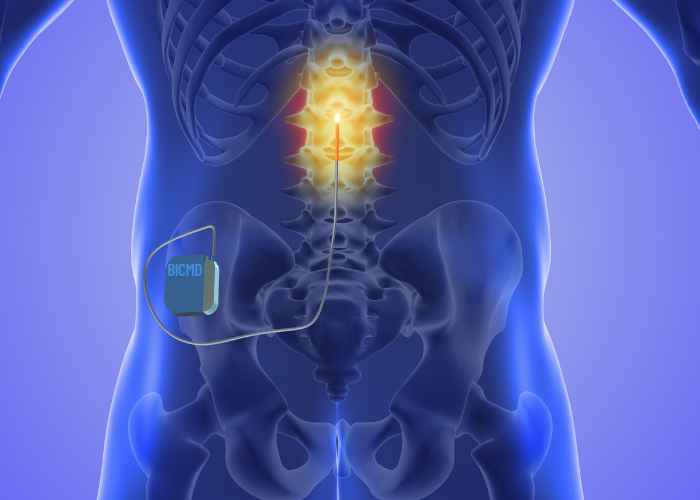
Spinal cord stimulation – If failed back surgery syndrome includes pain that radiates into the arms or legs, spinal cord manipulation may be an option. Spinal cord manipulation uses an implantable device to send gentle electrical pulses to the nerve roots that are causing pain. These electrical signals mask the pain signals and provide relief.

Pain-relieving medication – Some patients are best served by pain-relieving medications, including oral opioids or intrathecal pain-medicine delivery pumps.

Spinal cord injections – Epidural and facet joint injections are often used to treat lasting back pain.

Revision surgery – Depending on the cause of the failed back surgery syndrome, some surgeons will recommend a second surgery to repair the cause of the ongoing pain.

Suffering from failed back surgery syndrome?

For more information on failed back surgery syndrome, or to have your back pain evaluated by one of our board-certified orthopedic surgeons, please click on “Get Started” now. BICMD’s nationwide network of orthopedic surgeons can help you decide which treatment is best for your back pain. Our physicians are top orthopedic doctors hand-picked from reputable organizations nationwide and are extremely skilled with failed back surgery treatment options.

A spinal cord injury is a serious condition that requires expert care. Though there are no current treatments to reverse damage to an injured spinal cord, surgery is often required to prevent or minimize any further damage. The type and extent of the spinal cord injury dictates what kind of spinal surgery is the appropriate treatment.

For more information on Spinal Cord Injuries, or to have your injury evaluated by one of our orthopedic surgeons, please click on “Get Started” now. BICMD’s nationwide network of orthopedic surgeons can help you decide which treatment is best for you. Our physicians are top doctors hand-picked from reputable organizations nationwide and are extremely skilled with spinal cord injuries and possible treatment options.

This list contains some of our most commonly seen conditions, but it is by no means exhaustive. Our neck, back, and spine doctors handle the same wide variety of conditions that an in-person physician would.

No one should have to live with back and neck pain. Today’s advancements in orthopedic spine care have made it possible for many to live a pain-free life while maintaining full neck and back function.  A consultation with one of our neck, back, and spine experts is the first step on your path to recovery. Let us discuss the options that will work for you and your specific issue.  Our physicians are committed to helping you feel better.

Click on “Get Started” to reach one of our orthopedic telemedicine experts.

Best in Class Neck, Back & Spine Specialists

  • Federico Girardi, MD
    • Attending surgeon, top-ranked Hospital for Special Surgery
    • Professor of Orthopedic Surgery, Weill Cornell Medical College
  • Kathleen Davenport, MD
    • Director of physiatry, Hospital for Special Surgery Florida
    • Executive committee of the USA Task Force on Dancer Health

I've now made a decision about my next steps and the BICMD platform truly fulfilled its promise to provide expert second opinions.

-Alex Miller

The neck, back & spine advice you need to move forward